Tuesday, April 14, 2020

G protein-coupled receptor


G protein-coupled receptor


G protein-coupled receptor (GPCR), also called seven-transmembrane receptor or heptahelical receptor, protein located in the cell membrane that binds extracellular substances and transmits signals from these substances to an intracellular molecule called a G protein (guanine nucleotide-binding protein). GPCRs are found in the cell membranes of a wide range of organisms, including mammals, plants, microorganisms, and invertebrates. There are numerous different types of GPCRs—some 1,000 types are encoded by the human genome alone—and as a group they respond to a diverse range of substances, including light, hormones, amines, neurotransmitters, and lipids. Some examples of GPCRs include beta-adrenergic receptors, which bind epinephrine; prostaglandin E2 receptors, which bind inflammatory substances called prostaglandins; and rhodopsin, which contains a photoreactive chemical called retinal that responds to light signals received by rod cells in the eye. The existence of GPCRs was demonstrated in the 1970s by American physician and molecular biologist Robert J. Lefkowitz. Lefkowitz shared the 2012 Nobel Prize for Chemistry with his colleague Brian K. Kobilka, who helped to elucidate GPCR structure and function.
A GPCR is made up of a long protein that has three basic regions: an extracellular portion (the N-terminus), an intracellular portion (the C-terminus), and a middle segment containing seven transmembrane domains. Beginning at the N-terminus, this long protein winds up and down through the cell membrane, with the long middle segment traversing the membrane seven times in a serpentine pattern. The last of the seven domains is connected to the C-terminus. When a GPCR binds a ligand (a molecule that possesses an affinity for the receptor), the ligand triggers a conformational change in the seven-transmembrane region of the receptor. This activates the C-terminus, which then recruits a substance that in turn activates the G protein associated with the GPCR. Activation of the G protein initiates a series of intracellular reactions that end ultimately in the generation of some effect, such as increased heart rate in response to epinephrine or changes in vision in response to dim light (see second messenger).
Both inborn and acquired mutations in genes encoding GPCRs can give rise to disease in humans. For example, an inborn mutation of rhodopsin results in continuous activation of intracellular signaling molecules, which causes congenital night blindness. In addition, acquired mutations in certain GPCRs cause abnormal increases in receptor activity and expression in cell membranes, which can give rise to cancer. Because GPCRs play specific roles in human disease, they have provided useful targets for drug development. The antipsychotic agents clozapine and olanzapine block specific GPCRs that normally bind dopamine or serotonin. By blocking the receptors, these drugs disrupt the neural pathways that give rise to symptoms of schizophrenia. There also exist a variety of agents that stimulate GPCR activity. The drugs salmeterol and albuterol, which bind to and activate beta-adrenergic GPCRs, stimulate airway opening in the lungs and thus are used in the treatment of some respiratory conditions, including chronic obstructive pulmonary disease and asthma.

Wednesday, April 8, 2020

Lymphocytes: Definition, Ranges,Count & Functions


 Lymphocytes: Definition, Ranges,Count & Functions
 Lymphocytes: Definition, Ranges,Count & Functions 


Lymphocytes are one of several different types of white blood cells. Each type of white blood cell has a specific function, and they all work together to fight illness and disease.
White blood cells are an important part of your immune system. They help your body fight antigens, which are bacteria, viruses, and other toxins that make you sick. If your doctor says you have a weakened immune system, that means there aren’t enough white blood cells in your bloodstream.


 Lymphocytes: Definition, Ranges,Count & Functions
 Lymphocytes: Definition, Ranges,Count & Functions 


Lymphocyte levels can change according to a person’s race, gender, location, and lifestyle habits.
The normal lymphocyte range in adults is between 1,000 and 4,800 lymphocytes in 1 microliter (µL) of blood. In children, the normal range is between 3,000 and 9,500 lymphocytes in 1 µL of blood.
Unusually high or low lymphocyte counts can be a sign of disease.

 Lymphocytes: Definition, Ranges,Count & Functions
 Lymphocytes: Definition, Ranges,Count & Functions 

Lymphocyte Counts

Lymphocytes are a component of complete blood count (CBC) tests that include a white blood cell differential, in which the levels of the major types of white blood cells are measured. Such tests are used to assist in the detection, diagnosis, and monitoring of various medical conditions. Lymphocyte counts that are below the reference range, which varies for adults and children, may be indicative of lymphocytopenia (lymphopenia), whereas those above it are a sign of lymphocytosis. Lymphocytopenia is associated with a variety of conditions, ranging from malnutrition to rare inherited disorders such as ataxia-telangiectasia or severe combined immunodeficiency syndrome. Lymphocytosis typically is associated with infections, such as mononucleosis or whooping cough, certain cancers of the blood or lymphatic system such as multiple myeloma and chronic lymphocytic leukemia, and autoimmune disorders that cause chronic inflammation, such as inflammatory bowel disease.

A low lymphocyte count, called lymphocytopenia, usually occurs because:
  • your body isn’t producing enough lymphocytes
  • lymphocytes are being destroyed
  • lymphocytes are trapped in your spleen or lymph nodes
Lymphocytopenia can point to a number of conditions and diseases. Some, like the flu or mild infections, aren’t serious for most people. But a low lymphocyte count puts you at greater risk of infection.
Other conditions that can cause lymphocytopenia include:
  • undernutrition
  • HIV and AIDS
  • influenza
  • autoimmune conditions, such as lupus
  • some cancers, including lymphocytic anemia, lymphoma, and Hodgkin disease
  • steroid use
  • radiation therapy
  • certain drugs, including chemotherapy drugs
  • some inherited disorders, such as Wiskott-Aldrich syndrome and DiGeorge syndrome

Lymphocytosis, or a high lymphocyte count, is common if you’ve had an infection. High lymphocyte levels that persist may point to a more serious illness or disease, such as:
  • viral infections, including measles, mumps, and mononucleosis
  • adenovirus
  • hepatitis
  • influenza
  • tuberculosis
  • toxoplasmosis
  • cytomegalovirus
  • brucellosis
  • vasculitis
  • acute lymphocytic leukemia
  • chronic lymphocytic leukemia
  • HIV and AIDS

 Lymphocytes: Definition, Ranges,Count & Functions
 Lymphocytes: Definition, Ranges,Count & Functions 


There are different types of B cells and T cells that have specific roles in the body and the immune system.

B cells

Memory B cells circulate in the body to start a fast antibody response when they find a foreign substance. They remain in the body for decades and become memory cells, which remember previously found antigens and help the immune system respond faster to future attacks.
Regulatory B cells or Bregs make up around 0.5 percent of all B cells in healthy people. Although few in number, they have a vital role to play.
Bregs have protective anti-inflammatory effects in the body and stop lymphocytes that cause inflammation. They also interact with several other immune cells and promote the production of regulatory T cells or Tregs.

T cells

Killer or cytotoxic T cells scan the surface of cells in the body to see if they have become infected with germs, or if they have turned cancerous. If so, they kill these cells.
Helper T cells “help” other cells in the immune system to start and control the immune response against foreign substances.
There are different types of helper T cells, and some are more effective than others against different types of germs.
For instance, a Th1 cell is more effective against germs that cause infection inside other cells, such as bacteria and viruses, while a Th2 cell is more effective against germs that cause infection outside of cells, such as certain bacteria and parasites.
Tregs control or suppress other cells in the immune system. They have both helpful and harmful effects.
They maintain tolerance to germs, prevent autoimmune diseases, and limit inflammatory diseases. But they can also suppress the immune system from doing its job against certain antigens and tumors.
Memory T cells protect the body against previously found antigens. They live for a long time after an infection is over, helping the immune system to remember previous infections.
If the same germ enters the body a second time, memory T cells remember it and quickly multiply, helping the body to fight it more quickly.
Natural killer T cells are a mixed group of T cells that share characteristics of both T cells and natural killer cells. They can influence other immune cells and control immune responses against substances in the body that trigger an immune response.


Friday, April 3, 2020

Lotka-Volterra equations in Ecology


      Lotka-Volterra equations in Ecology

Lotka-Volterra equation


The effects of species interactions on the population dynamics of the species involved can be predicted by a pair of linked equations that were developed independently during the 1920s by American mathematician and physical scientist Alfred J. Lotka and Italian physicist Vito Volterra. Today the Lotka-Volterra equations are often used to assess the potential benefits or demise of one species involved in competition with another species:

dN1/dt = r1N1(1 – N1/K1 – α1,2N2/K2)dN2/dt = r2N2(1 – N2/K2 – α2,1N1/K1)
Here r = rate of increase, N = population size, and K = carrying capacity of any given species. In the first equation, the change in population size of species 1 over a specific period of time (dN1/dt) is determined by its own population dynamics in the absence of species 2 (r1N1[1 – N1/K1]) as well as by its interaction with species 2 (α1,2N2/K2). As the formula implies, the effect of species 2 on species 1 (α1,2) in turn is determined by the population size and carrying capacity of species 2 (N2 and K2).
Lotka-Volterra equation


The possible outcomes of interactions between two species are predicted on the basis of the relative strengths of self-regulation versus the species interaction term. For instance, species 2 will drive species 1 to local extinction if the term α1,2N2/K2 exceeds the term r1N1(1 − N1/K1)—though the term α1,2N2/K2 will exert a decreasing influence over the growth rate of species 1 as α1,2N2/K2 diminishes. Consequently, the first equation represents the amount by which the growth rate of species 1 over a specific time period will be reduced by its interaction with species 2. In the second equation, the obverse applies to the dynamics of species 2.
In the case of interspecific competition, if the effects of both species on each other are approximately equivalent with respect to the strength of self-regulation in each species, the populations of both species may stabilize; however, one species may gradually exclude the other over time. The competitive exclusion scenario is dependent on the initial population size of each species. For instance, when the interspecific effects of each species upon the abundance of its competitor are approximately equal, the species with the higher initial abundance is likely to drive the species with a lower initial abundance to exclusion.

Lotka-Volterra equation


The basic equations given above, describing the dynamics deriving from an interaction between two competitors, have undergone several modifications. Chief among these modifications is the development of a subset of Lotka-Volterra equations that calculate the effects of interacting predator and prey populations. In their simplest forms, these modified equations bear a strong resemblance to the equations above, which are used to assess competition between two species:

dNprey/dt = rprey × Nprey(1 − Nprey/Kprey – αprey, pred × Npred/Kpred)dNpred/dt = rpred × Npred(1 − Npred/Kpred + αpred, prey × Nprey/Kprey)


Lotka-Volterra equation


Here the terms Npred and Kpred denote the size of the predator population and its carrying capacity. Similarly, the population size and carrying capacity of the prey species are denoted by the terms Nprey and Kprey, respectively. The coefficient αprey, pred represents the reduction in the growth rate of prey species due to its interaction with the predator, whereas αpred, prey represents the increase in growth rate of the predator population due to its interaction with prey population.

Several additional modifications to the Lotka-Volterra equations are possible, many of which have focused on the incorporation of influences of spatial refugia (predator-free areas) from predation on prey dynamics.

Thursday, April 2, 2020

Metapopulation in Ecology


               Metapopulation in Ecology 


Metapopulation in Ecology
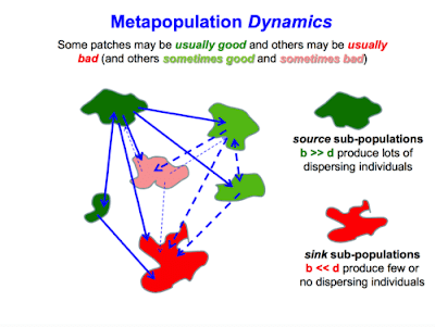
Metapopulation Dynamics

Metapopulation, in ecology, a regional group of connected populations of a species. For a given species, each metapopulation is continually being modified by increases (births and immigrations) and decreases (deaths and emigrations) of individuals, as well as by the emergence and dissolution of local populations contained within it. As local populations of a given species fluctuate in size,theybecome vulnerable to extinction
during periods when their numbers are low. Extinction of local populations is common in some species, and the regional persistence of such species is dependent on the existence of a metapopulation. Hence, elimination of much of the metapopulation structure of some species can increase the chance of regional extinction of species.


Metapopulation in Ecology
Metapopulation Structure 

The structure of metapopulations varies among species. In some species one population may be particularly stable over time and act as the source of recruits into other, less stable populations. For example, populations of the checkerspot butterfly (Euphydryas editha) in California have a metapopulation structure consisting of a number of small satellite populations that surround a large source population on which they rely for new recruits. The satellite populations are too small and fluctuate too much to maintain themselves indefinitely. Elimination of the source population from this metapopulation would probably result in the eventual extinction of the smaller satellite populations.


Metapopulation in Ecology
Metapopulation in Ecology 


In other species, metapopulations may have a shifting source. Any one local population may temporarily be the stable source population that provides recruits to the more unstable surrounding populations. As conditions change, the source population may become unstable, as when disease increases locally or the physical environment deteriorates. Meanwhile, conditions in another population that had previously been unstable might improve, allowing this population to provide recruits.

Featured Post

6 Surprising Ways Garlic Boosts Your Health

6 Surprising Ways Garlic Boosts Your Health : 6 Surprising Ways Garlic Boosts Your Health Garlic. Roasted in olive oil, it can ...