Showing posts with label Cell biology. Show all posts

Tuesday, April 14, 2020

G protein-coupled receptor


G protein-coupled receptor


G protein-coupled receptor (GPCR), also called seven-transmembrane receptor or heptahelical receptor, protein located in the cell membrane that binds extracellular substances and transmits signals from these substances to an intracellular molecule called a G protein (guanine nucleotide-binding protein). GPCRs are found in the cell membranes of a wide range of organisms, including mammals, plants, microorganisms, and invertebrates. There are numerous different types of GPCRs—some 1,000 types are encoded by the human genome alone—and as a group they respond to a diverse range of substances, including light, hormones, amines, neurotransmitters, and lipids. Some examples of GPCRs include beta-adrenergic receptors, which bind epinephrine; prostaglandin E2 receptors, which bind inflammatory substances called prostaglandins; and rhodopsin, which contains a photoreactive chemical called retinal that responds to light signals received by rod cells in the eye. The existence of GPCRs was demonstrated in the 1970s by American physician and molecular biologist Robert J. Lefkowitz. Lefkowitz shared the 2012 Nobel Prize for Chemistry with his colleague Brian K. Kobilka, who helped to elucidate GPCR structure and function.
A GPCR is made up of a long protein that has three basic regions: an extracellular portion (the N-terminus), an intracellular portion (the C-terminus), and a middle segment containing seven transmembrane domains. Beginning at the N-terminus, this long protein winds up and down through the cell membrane, with the long middle segment traversing the membrane seven times in a serpentine pattern. The last of the seven domains is connected to the C-terminus. When a GPCR binds a ligand (a molecule that possesses an affinity for the receptor), the ligand triggers a conformational change in the seven-transmembrane region of the receptor. This activates the C-terminus, which then recruits a substance that in turn activates the G protein associated with the GPCR. Activation of the G protein initiates a series of intracellular reactions that end ultimately in the generation of some effect, such as increased heart rate in response to epinephrine or changes in vision in response to dim light (see second messenger).
Both inborn and acquired mutations in genes encoding GPCRs can give rise to disease in humans. For example, an inborn mutation of rhodopsin results in continuous activation of intracellular signaling molecules, which causes congenital night blindness. In addition, acquired mutations in certain GPCRs cause abnormal increases in receptor activity and expression in cell membranes, which can give rise to cancer. Because GPCRs play specific roles in human disease, they have provided useful targets for drug development. The antipsychotic agents clozapine and olanzapine block specific GPCRs that normally bind dopamine or serotonin. By blocking the receptors, these drugs disrupt the neural pathways that give rise to symptoms of schizophrenia. There also exist a variety of agents that stimulate GPCR activity. The drugs salmeterol and albuterol, which bind to and activate beta-adrenergic GPCRs, stimulate airway opening in the lungs and thus are used in the treatment of some respiratory conditions, including chronic obstructive pulmonary disease and asthma.

Thursday, March 19, 2020

Cancer comes from overproduction and malfunction of the body's own cells

Cancer

Cancer comes from overproduction and         malfunction of the body's own cells.


  • Cancer is the uncontrolled growth of abnormal cells anywhere in a body.
  • There are over 200 types of cancer.
  • Anything that may cause a normal body cell to develop abnormally potentially can cause cancer; general categories of cancer-related or causative agents are as follows: chemical or toxic compound exposures, ionizing radiation, some pathogens, and human genetics.
  • Cancer symptoms and signs depend on the specific type and grade of cancer; although general signs and symptoms are not very specific the following can be found in patients with different cancers: fatigueweight losspain, skin changes, change in bowel or bladder function, unusual bleeding, persistent cough or voice change, fever, lumps, or tissue masses.
  • Although there are many tests to screen and presumptively diagnose cancer, the definite diagnosis is made by examination of a biopsy sample of suspected cancer tissue.
  • Cancer staging is often determined by biopsy results and helps determine the cancer type and the extent of cancer spread; staging also helps caregivers determine treatment protocols. In general, in most staging methods, the higher the number assigned (usually between 0 to 4), the more aggressive the cancer type or more widespread is the cancer in the body. Staging methods differ from cancer to cancer and need to be individually discussed with your health care provider.
  • Treatment protocols vary according to the type and stage of the cancer. Most treatment protocols are designed to fit the individual patient's disease. However, most treatments include at least one of the following and may include all: surgery, chemotherapy, and radiation therapy.
  • There are many listed home remedies and alternative treatments for cancers but patients are strongly recommended to discuss these before use with their cancer doctors.
  • The prognosis of cancer can range from excellent to poor. The prognosis depends on the cancer type and its staging with those cancers known to be aggressive and those staged with higher numbers (3 to 4) often have a prognosis that ranges more toward poor. 
  • What is cancer? 
  • Cancer is the uncontrolled growth of abnormal cells anywhere in a body. These abnormal cells are termed cancer cells, malignant cells, or tumor cells. These cells can infiltrate normal body tissues. Many cancers and the abnormal cells that compose the cancer tissue are further identified by the name of the tissue that the abnormal cells originated from (for example, breast cancer, lung cancercolorectal cancer)Cancer is not confined to humans; animals and other living organisms can get cancer. Below is a schematic that shows normal cell division and how when a cell is damaged or altered without repair to its system, the cell usually dies. Also shown is what occurs when such damaged or unrepaired cells do not die and become cancer cells and show uncontrolled division and growth -- a mass of cancer cells develop. Frequently, cancer cells can break away from this original mass of cells, travel through the blood and lymph systems, and lodge in other organs where they can again repeat the uncontrolled growth cycle. This process of cancer cells leaving an area and growing in another body area is termed metastatic spread or metastasis. For example, if breast cancer cells spread to a bone, it means that the individual has metastatic breast cancer to bone. This is not the same as "Bone cancer" which would mean the cancer had started in the bone.
    The following table (National Cancer Institute 2016) gives the estimated numbers of new cases and deaths for each common cancer type:
  • Cancer TypeEstimated New CasesEstimated Deaths
    Bladder76,96016,390
    Breast (Female -- Male)246,660 -- 2,60040,450 -- 440
    Colorectal Cancer134,49049,190
    Endometrial60,05010,470
    Kidney (Renal Cell and Renal Pelvis) Cancer62,70014,240
    Leukemia (All Types)60,14024,400
    Lung (Including Bronchus)224,390158,080
    Melanoma76,38010,130
    Non-Hodgkin Lymphoma72,58020,150
    Pancreatic53,07041,780
    Prostate180,89026,120
    Thyroid64,3001,980
  • The three most common cancers in men, women, and children in the U.S. are as follows:
    The incidence of cancer and cancer types are influenced by many factors such as age, gender, race, local environmental factors, diet, and genetics. Consequently, the incidence of cancer and cancer types vary depending on these variable factors. For example, the World Health Organization (WHO) provides the following general information about cancer worldwide:
    • Cancer is a leading cause of death worldwide. It accounted for 8.2 million deaths (around 22% of all deaths not related to communicable diseases; most recent data from WHO).
    • Lung, stomach, liver, colon, and breast cancer cause the most cancer deaths each year.
    • Deaths from cancer worldwide are projected to continue rising, with an estimated 13.1 million deaths in 2030 (about a 70% increase).
    Different areas of the world may have cancers that are either more or less predominant then those found in the U.S. One example is that stomach cancer is often found in Japan, while it is rarely found in the U.S. This usually represents a combination of environmental and genetic factors.
    The objective of this article is to introduce the reader to general aspects of cancers. It is designed to be an overview of cancer and cannot cover every cancer type. This article will also attempt to help guide the reader to more detailed sources about specific cancer types.
  •  What are risk factors and causes of cancer?

  • Anything that may cause a normal body cell to develop abnormally potentially can cause cancer. Many things can cause cell abnormalities and have been linked to cancer development. Some cancer causes remain unknown while other cancers have environmental or lifestyle triggers or may develop from more than one known cause. Some may be developmentally influenced by a person's genetic makeup. Many patients develop cancer due to a combination of these factors. Although it is often difficult or impossible to determine the initiating event(s) that cause a cancer to develop in a specific person, research has provided clinicians with a number of likely causes that alone or in concert with other causes, are the likely candidates for initiating cancer. The following is a listing of major causes and is not all-inclusive as specific causes are routinely added as research advances:
    Chemical or toxic compound exposures: Benzene, asbestos, nickel, cadmium, vinyl chloride, benzidine, N-nitrosamines, tobacco or cigarette smoke (contains at least 66 known potential carcinogenic chemicals and toxins), asbestos, and aflatoxin
    Ionizing radiation: Uranium, radon, ultraviolet rays from sunlight, radiation from alpha, beta, gamma, and X-ray-emitting sources
    Pathogens: Human papillomavirus (HPV), EBV or Epstein-Barr virus, hepatitis virusesB and C, Kaposi's sarcoma-associated herpes virus (KSHV), Merkel cell polyomavirus, Schistosoma spp., and Helicobacter pylori; other bacteria are being researched as possible agents.
  • Genetics: A number of specific cancers have been linked to human genes and are as follows: breast, ovarian, colorectal, prostate, skin and melanoma; the specific genes and other details are beyond the scope of this general article so the reader is referred to the National Cancer Institute for more details about genetics and cancer.
    It is important to point out that most everyone has risk factors for cancer and is exposed to cancer-causing substances (for example, sunlight, secondary cigarette smoke, and X-rays) during their lifetime, but many individuals do not develop cancer. In addition, many people have the genes that are linked to cancer but do not develop it. Why? Although researchers may not be able give a satisfactory answer for every individual, it is clear that the higher the amount or level of cancer-causing materials a person is exposed to, the higher the chance the person will develop cancer. In addition, the people with genetic links to cancer may not develop it for similar reasons (lack of enough stimulus to make the genes function). In addition, some people may have a heightened immune response that controls or eliminates cells that are or potentially may become cancer cells. There is evidence that even certain dietary lifestyles may play a significant role in conjunction with the immune system to allow or prevent cancer cell survival. For these reasons, it is difficult to assign a specific cause of cancer to many individuals.
    Recently, other risk factors have been added to the list of items that may increase cancer risk. Specifically, red meat (such as beef, lamb, and pork) was classified by the International Agency for Research on Cancer as a high-risk agent for potentially causing cancers; in addition processed meats (salted, smoked, preserved, and/or cured meats) were placed on the carcinogenic list. Individuals that eat a lot of barbecued meat may also increase risk due to compounds formed at high temperatures. Other less defined situations that may increase the risk of certain cancers include obesity, lack of exercise, chronic inflammation, and hormones, especially those hormones used for replacement therapy. Other items such as cell phones have been heavily studied. In 2011, the World Health Organization classified cell phone low energy radiation as "possibly carcinogenic," but this is a very low risk level that puts cell phones at the same risk as caffeine and pickled vegetables.
  • Proving that a substance does not cause or is not related to increased cancer risk is difficult. For example, antiperspirants are considered to possibly be related to breast cancer by some investigators and not by others. The official stance by the NCI is "additional research is needed to investigate this relationship and other factors that may be involved." This unsatisfying conclusion is presented because the data collected so far is contradictory. Other claims that are similar require intense and expensive research that may never be done. Reasonable advice might be to avoid large amounts of any compounds even remotely linked to cancer, although it may be difficult to do in complex, technologically advanced modern societies.
  • What are cancer symptoms and signs?


  • Symptoms and signs of cancer depend on the type of cancer, where it is located, and/or where the cancer cells have spread. For example, breast cancer may present as a lump in the breast or as nipple discharge while metastatic breast cancer may present with symptoms of pain (if spread to bones), extreme fatigue (lungs), or seizures (brain). A few patients show no signs or symptoms until the cancer is far advanced.

    The American Cancer Society describes seven warning signs and/or symptoms that a cancer may be present, and which should prompt a person to seek medical attention. The word CAUTION can help you remember these.
    • Change in bowel or bladder habits
    • sore throat that does not heal
    • Unusual bleeding or discharge (for example, nipple secretions or a "sore" that will not heal that oozes material)
    • Thickening or lump in the breast, testicles, or elsewhere
    • Indigestion (usually chronic) or difficulty swallowing
    • Obvious change in the size, color, shape, or thickness of a wart or mole
    • Nagging cough or hoarseness
    Other signs or symptoms may also alert you or your doctor to the possibility of your having some form of cancer. These include the following:
    • Unexplained loss of weight or loss of appetite
    • A new type of pain in the bones or other parts of the body that may be steadily worsening, or come and go, but is unlike previous pains one has had before
    • Persistent fatigue, nausea, or vomiting
    • Unexplained low-grade fevers with may be either persistent or come and go
    • Recurring infections which will not clear with usual treatment. 
    • Anyone with these signs and symptoms should consult their doctor; these symptoms may also arise from noncancerous conditions.
      Many cancers will present with some of the above general symptoms but often have one or more symptoms that are more specific for the cancer type. For example, lung cancer may present with common symptoms of pain, but usually the pain is located in the chest. The patient may have unusual bleeding, but the bleeding usually occurs when the patient coughs. Lung cancer patients often become short of breath and then become very fatigued.
      Because there are so many cancer types (see next section) with so many nonspecific and sometimes more specific symptoms, the best way to learn about signs and symptoms of specific cancer types is to spend a few moments researching symptoms of a specific body area in question. Conversely, a specific body area can be searched to discover what signs and symptoms a person should look for in that area that is suspected of having cancer. The following examples are two ways to proceed to get information on symptoms:
      • Use a search engine (Google, Bing) to find links to cancer by listing the symptom followed by the term "cancer" or if you know the type you want information about, (lung, brain, breast) use MedicineNet’s search option. For example, listing "blood in urine and cancer" will bring a person to web sites that list possible organs and body systems where cancer may produce the listed symptoms.
      • Use a search engine as above and list the suspected body area and cancer (for example, bladder and cancer), and the person will see sites that list the signs and symptoms of cancer in that area (blood in urine being one of several symptoms listed).
      • Be aware that many web sites are not necessarily reviewed by a health care professional and could contain information that is not accurate. Your health care professional is ultimately the best resource if you have concerns.
      • In addition, if the cancer type is known (diagnosed), then even more specific searches can be done listing the diagnosed cancer type and whatever may be questioned about the cancer (symptoms, tumor grades, treatments, prognosis, and many other items).
        One's own research should not replace consulting a health care provider if someone is concerned about cancer.
      • What are the different types of cancer?


        • Carcinoma: Cancer that begins in the skin or in tissues that line or cover internal organs -- "skin, lung, colon, pancreatic, ovarian cancers," epithelial, squamous and basal cell carcinomas, melanomas, papillomas, and adenomas
        • Sarcoma: Cancer that begins in bone, cartilage, fat, muscle, blood vessels, or other connective or supportive tissue -- "bone, soft tissue cancers," osteosarcoma, synovial sarcoma, liposarcoma, angiosarcoma, rhabdosarcoma, and fibrosarcoma
        • Leukemia: Cancer that starts in blood-forming tissue such as the bone marrow and causes large numbers of abnormal blood cells to be produced and enter the blood -- "leukemia," lymphoblastic leukemias (ALL and CLL), myelogenous leukemias (AML and CML), T-cell leukemia, and hairy-cell leukemia
        • Lymphoma and myeloma: Cancers that begin in the cells of the immune system -- "lymphoma," T-cell lymphomas, B-cell lymphomas, Hodgkin lymphomas, non-Hodgkin lymphoma, and lymphoproliferative lymphomas
        • Central nervous system cancers: Cancers that begin in the tissues of the brain and spinal cord -- "brain and spinal cord tumors," gliomas, meningiomas, pituitary adenomas, vestibular schwannomas, primary CNS lymphomas, and primitive neuroectodermal tumors. 
        • What specialists treat cancer?


        • A doctor who specializes in the treatment of cancer is called an oncologist. He or she may be a surgeon, a specialist in radiation therapy, or a medical oncologist. The first uses surgery to treat the cancer; the second, radiation therapy; the third, chemotherapy and related treatments. Each may consult with the others to develop a treatment plan for the particular patient.

          In addition, other specialists may be involved depending upon where the cancer is located. For example, ob-gyn specialists may be involved with uterine cancer while an immunologist maybe involved in treatment of cancers that occur in the immune system. Your primary care physician and main oncologist will help you to determine what specialists are best to be members of your treatment team.
        • How do health care professionals diagnose cancer?


        • Some cancers are diagnosed during routine screening examinations. These are usually tests that are routinely done at a certain age. Many cancers are discovered when you present to your health care professional with specific symptoms.

          A physical exam and medical history, especially the history of symptoms, are the first steps in diagnosing cancer. In many instances, the medical caregiver will order a number of tests, most of which will be determined by the type of cancer and where it is suspected to be located in or on the person's body. In addition, most caregivers will order a complete blood count, electrolyte levels and, in some cases, other blood studies that may give additional information.

          Imaging studies are commonly used to help physicians detect abnormalities in the body that may be cancer. X-rays, CT and MRI scans, and ultrasound are common tools used to examine the body. Other tests such as endoscopy, which with variations in the equipment used, can allow visualization of tissues in the intestinal tract, throat, and bronchi that may be cancerous. In areas that cannot be well visualized (inside bones or some lymph nodes, for example), radionuclide scanning is often used. The test involves ingestion or IV injection of a weakly radioactive substance that can be concentrated and detected in abnormal tissue.
        • The preceding tests can be very good at localizing abnormalities in the body; many clinicians consider that some of the tests provide presumptive evidence for the diagnosis of cancer. However, in virtually all patients, the definitive diagnosis of cancer is based on the examination of a tissue sample taken in a procedure called a biopsy from the tissue that may be cancerous, and then analyzed by a pathologist. Some biopsy samples are relatively simple to procure (for example, skin biopsy or intestinal tissue biopsy done with a device called an endoscope equipped with a biopsy attachment). Other biopsies may require as little as a carefully guided needle, or as much as a surgery (for example, brain tissue or lymph node biopsy). In some instances, the surgery to diagnose the cancer may result in a cure if all of the cancerous tissue is removed at the time of biopsy.
        • The biopsy can provide more than the definitive diagnosis of cancer; it can identify the cancer type (for example, the type of tissue found may indicate that the sample is from a primary [started there] or metastatic type of brain cancer [spread from another primary tumararising elsewhere in the body]) and thereby help to stage the cancer. The stage, or cancer staging, is a way for clinicians and researchers to estimate how extensive the cancer is in the patient's body.
          Is the cancer that has been found localized to its site of origin, or is it spread from that site to other tissues? A localized cancer is said to be at an early stage, while one which has spread is at and advanced stage. The following section describes the general staging methods for cancers.

Wednesday, March 18, 2020

Protoplast Isolation and Culture

Plant Protoplast Culture: Meaning, History and Principles


Let us make an in-depth study of the meaning, history and principles of plant protoplast culture.


What is a Protoplast?

It is known that each and every plant cell possesses a definite cellulosic cell wall and the protoplast lies within the cell wall except some reproductive cells and the free floating cells in some fruit juices like coconut water.

Therefore, protoplast of plant cell consists of plasma-lemma and everything contained within it.
But those of importance to plant protoplast culture are pro­duced experimentally by the removal of cell wall by either enzymatically or mechanical means from the artificially plasmolysed plant cells. Ex­perimentally produced protoplasts are known as isolated protoplasts.
According to Torrey and Landgren (1977) “the isolated protoplasts are the cells with their walls stripped off and removed from the prox­imity of their neighbouring cells”. Vasil (1980) defines that “the protoplast is a part of plant cell which lies within the cell wall and can be plas­molysed and which can be isolated by removing the cell wall by mechanical or enzymatic proce­dure”. Therefore, isolated protoplast is only a naked plant cell surrounded by plasma membra­ne—which is potentially capable of cell wall re­generation, cell division, growth and plant regen­eration in culture.

Brief Past History:

J. Klercker (1892):
First isolated proto­plast mechanically from plasmolyzed cell of wa­ter warrior (Stratiotes aloides). No attempt was made to culture them.
E. Kiister (1927):
In the fruits of several plants like Solatium nigram, Lycopersicon esculenium etc. the cell wall are hydrolysed during fruits ripening process so that free protoplasts and protoplasmic units are left. Kuster preferred such physiological method for isolating proto­plasts. No report of culture was available.

R. Chambers and K. Hofler (1931):
Were able to isolate few protoplasts by using thin slices of epidermis of onion bulb immersed in 1M sucrose until the protoplast shrunk away from their enclosing walls and then cutting sheets of epidermis with a sharp knife. Report of culture was not available.

E. C. Cocking (I960):
First reported the enzymatic method for isolation of protoplast in a large number from root tip cells of Lycopersicon esculentum by using a concentrated solution of cellulase enzyme, prepared from cultures of the fungus Myrothecium verrucaria to degrade cell wall.
I. Takebe, Y. Otsuki and S. Aoki (1968):
First employed the commercial preparation of cellulase and macerozyme sequentially (in two steps) for the isolation of mesophyll protoplast of tobacco.
J. B. Power and E. C. Cocking (1968):


Demonstrated first that the mixture of such two enzymes (cellulase + macerozyme) can be used simultaneously (one step method) for the isola­tion of protoplasts.

I. Takebe, G. Labib, G. Melchers (1971):
First reported the plant regeneration from isolated protoplast in Nicotiana tabacum.
P. S. Carlson, H. H. Smith, R. D. Dearing (1972):
First reported a somatic hy­brid in higher plants involving two different sex­ually compatible species of mesophyll protoplast (N. glauca x N. langsdorffi).

Different Sources of Plant Tissue and Their Condition for Protoplast Isolation:

Protoplast can be isolated either directly from the different parts of whole plant or indi­rectly from in vitro cultured tissue. Convenient and suitable materials are leaf, mesophyll and cells from liquid suspension cul­tures. Protoplast yield and viability are pro­foundly influenced by the growing conditions of plants serving leaf mesophyll sources.
The age of the plant and of the leaf and the prevailing conditions of light, photoperiod, humidity, tem­perature, nutrition and watering are contribut­ing factors. Cell suspension cultures may provide a more reliable source for obtaining consistent qua­lity protoplasts. It is necessary, however, to es­tablish and maintain the cells at maximum growth rates and utilize the cell at the early log phase.

Principles of Protoplast Culture:


The basic principle of protoplast culture is the aseptic isolation of large number of intact living protoplasts removing their cell wall and cultures them on a suitable nutrient medium for their requisite growth and development. Protoplast can be isolated from varieties of plant tissues. Convenient and suitable materials are leaf mesophyll and cells from liquid suspension culture. Protoplast yield and viabil­ity are greatly influenced by the growing condi­tion of the plant as well as the cells.

The essential step of the isolation of proto­plast is the removal of the cell wall without dam­aging the cell or protoplasts. The plant cell is an osmotic system. The cell wall exerts the inward pressure upon the enclosed protoplasts. Like­wise, the protoplast also puts equal and oppo­site pressure upon the cell wall. Thus, both the pressures are balanced.

Now if the cell wall is re­moved, the balanced pressures will be disturbed. As a result, the outward pressure of protoplast will be greater and at the same time in absence of cell wall, irresistible expansion of protoplast takes place due to huge inflow of water from the external medium. Greater outward pressure and the expansion of protoplast cause it to burst.

So, the isolated protoplast is an osmotically frag­ile structure at its nascent stage. Therefore, if the cell wall is to be removed to isolate proto­plast, the cell or tissue must be placed in a hy­pertonic solution of a metabolically inert sugar such as mannitol at higher concentration (13%) to plasmolysis the cell away from the cell wall (Fig 12.2).

Mannitol, an alcoholic sugar, is easily transported across the plasmodesmata, provides a stable osmotic environment for the protoplasts and prevents the usual expansion and bursting of protoplast even after loss of cell wall. That is why, this hypertonic solution is known as os­motic stabilizer or plasmolyticum or osmolyticum.

Once the cells are stabilized in such a man­ner by plasmolysis the protoplasts are released from the containing cell wall either mechanically or enzymatically. Mechanical isolation (Fig 12.3) involves breaking open each cell compartment to liberate the protoplast. This operation can be done carefully on small pieces of tissue un­der a microscope using a micro-scalpel.


But very few protoplasts are obtained for a lot of time and effort. Large-scale attempts at mechani­cal isolation involves the disrupting tissue with fine stainless steel-bristled brush. This process may liberate more protoplasts with less efforts, but the percentage of yield of intact protoplasts is still very low.

A considerably more efficient way of liberating the protoplasts is to digest the cell walls away around them, using cell wall de­grading enzymes such as cellulase, hemicellulose, pectinase or macerozyme etc. These enzymes are isolated from fungi and available commercially (Table 12.1).


Period of treatment and concentration of enzymes are the critical factors and both factors should be standardized for particular plant tis­sue. Intact tissue can be incubated with a pecti­nase or macerozyme solution which will dissolve the middle lamella between the cells and so sep­arate them.

Subsequent treatment with cellu­lase will digest away the cellulosic layer of the cell wall. This process is known as sequential enzyme treatment or two step method as op­posed to a mixed enzyme treatment (one step method) in which both cellulase and pectinase or macerozyme are mixed so that the entire wall is broken down in a single operation (Fig. 12.4).

The isolated protoplasts can be cultured ei­ther static liquid or agarified medium. The pro­toplast media consist of mineral salts, vitamins, carbon sources and plant growth hormones as well as osmotic stabilizers and possibly organic nitrogen sources, coconut milk and organic acids.


In culture protoplast can reform a new cell wall around them. Once the wall is formed, the proto­plast becomes a cell. The cells from protoplasts subsequently enter cell division which is followed by the formation of callus and cell cultures. Such callus also retain the capacity for morphogenesis and plant regeneration. A brief list of plant regeneration from plant protoplast culture is given below (Table 12.2).


Tuesday, March 17, 2020

Molecular and Physiological Mechanisms of Membrane Receptor Systems


           Fig :Receptor in Secreting Cell

In cell biologyreceptors are special structures that can be found in cell membranes. These are made of protein molecules such as glycoproteins. Receptors bind (attach) to specialised molecules. If the receptor has this molecule, it is activated, but if it does not it is deactivated. Depending on its state, a change inside the cell happens.

Cell surface receptors (membrane receptors, transmembrane receptors) take part in communication between the cell and the outside world. Extracellular signalling molecules (usually hormonesneurotransmitterscytokinesgrowth factors or cell recognition molecules) attach to the receptor. This triggers changes in the function of the cell. The process is called signal transduction: The binding starts a chemical change on the inside of the membrane.


               Fig : Receptor in Target Cell

In short, receptors work like locks and keys. With the key, the lock can be locked or unlocked. If it is unlocked, the door belonging to it can be opened.

Structure
Part of the receptor sticks out of the cell membrane. The same applies to the membranes of cell organelles. A receptor's main function is to recognize and respond to a specific ligand, for example, a neurotransmitter or hormone. Some receptors respond to changes in 'transmembrane potential' (the difference in electric potential between the inside and the outside of a cell).


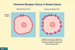
       Fig: Receptor status in Breast Cancer

The middle part, inside the membrane itself, is a protein-lined pore through the membrane, or 'ion channel'. When the ligand binds to the surface, the pore becomes accessible to ions, which then pass through. In other cases, where differences in electric potential occur, the receptor changes in shape, which causes change inside the cell.


 Fig: Hormone - Receptor Complex Enters        Nucleus 

The inside (or cytoplasmic) part of the receptor interacts with the inside of the cell or organelle. There are several different kinds of receptor, each of which acts in a different way.

Featured Post

6 Surprising Ways Garlic Boosts Your Health

6 Surprising Ways Garlic Boosts Your Health : 6 Surprising Ways Garlic Boosts Your Health Garlic. Roasted in olive oil, it can ...